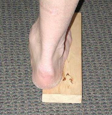
Pes cavus deformity
Pes cavus or high arched foot is characterized by increased medial longitudinal arch and rearfoot varus. It is also called inverted foot. In athletic activities, shockabsorption is a important role of feet by reducing high impact forces. High arch foot is commonly rigid and not flexible, it reduces shock absorping function of the foot. It leads to local and global injuries. Proper assessment of feet can give more information about distant injuries. Biomechanical assessment and correction through corrective exercises and orthotics/shoe modifications help athletic performance and also prevent recurrent injuries. High arch foot is more prone to inversion ankle sprain.
Causes
- Common in neurological disorders
- Involved structures are:
- Achilles tendon (tightness)
- Peroneus longus (tightness)
- Subtalar joint (inversion)
- First metatarsal (flexion)
Special tests
1. Wet paper test
Patient keep the foot on the paper with wet foot. It creates impression of foot on the paper. In cavus foot, medial part of the foot will not contact the ground. So, we can see small breadth of mid foot in wet paper.
2. Peek a boo sign
It is visible varus deformity of subtalar joint. There is loss of calcaneovalgus during weight bearing.
3. Coleman block test
This test is used to isolate the problem from first metatarsal. If coleman block test is positive, the condition is due to first metatarsal flexion. In this test, patient/athlete is asked to stand on the block with half lateral foot. So, medial part of the foot (first metatarsal) will free to flex downward. This is positive sign of coleman block test. Also therapist can see the loss of calcaneal varus during the test.

4. Isolating tight structures
Peroneus longus
Patient should be in standing position. Keep the therapist thumbs under the balls of the foot. one thumb is under first MTP joint and another thumb is under the lateral 4 MTP joints. Ask the patient to do plantarflexion. If peroneal longus is involved, The thumb under first MTP will get more pressure than the another thumb.
Achilles tendon
By checking calf flexibility in knee extended position helps to isolate gastronemeus tightness. Tight gastronemeus can cause external rotation of the foot in pes cavus.
Associated pathologies
- Overload calluses
- Stress fracture of 4th 5th metatarsal, Navicular, Tibia or Medial malleolus.
- Peroneal tendinitis, subluxation or dislocation.
- Hallux sesmoiditis
- Tight achilles and plantar fascia.
- Ankle instability and recurrent sprain.
- Ankle arthritis
- Knee pathology (Due to foot and tibial external rotation) - mainly lateral knee structures.
SHARE:


- Andrew K. Buldt, Pazit Levinger., Foot posture is associated with kinematics of the foot during gait: A comparison of normal, planus and cavus feet. (2015)
- Sarang N.Desai, Randolph Grierson, Arthur ManoliII., The Cavus Foot in Athletes: Fundamentals of Examination and Treatment. (2010)
| Name | : | Deva senathipathi |
| Qualifications | : | Physiotherapist |


