Radiography (x-ray)
- Introduction
- Parts of X-ray machine
- Views of X-ray
- Parameters
- X-ray absorption and object density
- Mechanism
Introduction
X-rays are electromagnetic waves produced in x-ray tube. Healthcare persons should call it as films, images or radiographs, but not x-ray. Direction of X-ray beam determines radiographic views include AP, PA, Oblique and Lateral.
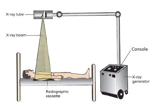
Parts of x-ray machine
- Operating console
It provides controls for radiology technician to adjust machine before taking x-ray.
- High frequency generator
It powers the x-ray tube for generating x-ray.
- X-ray tube
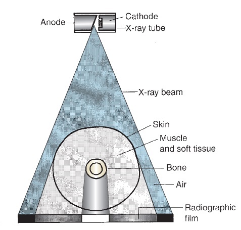
It is vacuum diode which consists cathode and anode. Electron passes from cathode to anode produces electromagnetic wave of x-ray. This x-ray beam passes into the body and then to the cassette.
- Radiographic cassette
It is placed in one side of the body and x-ray tube is position another side of the body, like in the image. Cassette holds x-ray film. It receives x-ray waves that were not absorbed by the body parts.
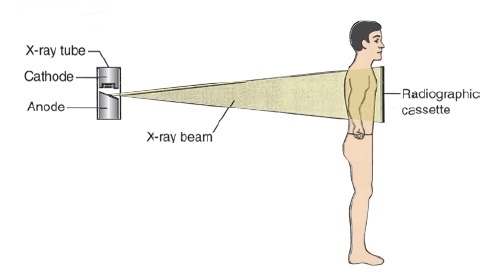
Views of x-ray
- PA view
X-ray beam travels from posterior to anterior as it traverses anatomic site. Example, chest x-ray PA view – x-ray tube is placed posterior side of the chest and cassette is placed in front of the chest.
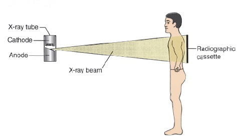
- AP view
X-ray beam passes through anatomic site from anterior to posterior. Portable X-ray machines use AP view. It is commonly used when the patient cant shifted to radiology department. Example, AP view of chest x-ray – The tube is in front of the chest and the cassette is placed in back side of the chest.
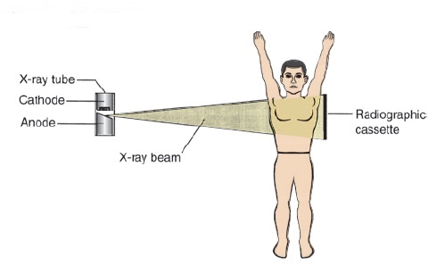
- Lateral view
X-ray beam travels from side to side. In this method, the x-ray tube will be on one side and the cassette will be on another side.
Parameters
- Tube potential (kVp)
Power and strength of x-ray beam (quality of x-ray).
- Tube intensity (Ma)
Number of photons generated in x-ray beam (quantity of x-ray)
- Exposurre time (S)
Time duration for preset x-ray beam.
- Focus to detector distance (Cm)
X-ray absorption and object density
X-Ray absorption is directly proportion with density of object. When object’s density increases, X-ray absorption will increase results in white appearance in the x-ray film.
There are black, grey and white colors in x-ray film and these are determined by objects density. Following are the density and color of object in the film,
- Air – black
- Fat – black (slightly less black than the air)
- Bone – white
- Metal – white
- Calcium – white
- Organs, muscles, tissues – shades of grey.
Mechanism
X-ray beam passed through the anatomic structure, some of rays absorbed in the structures (will not reach the x-ray cassette) will appear white and non-absorbed rays reach the cassette will appear as black in color. In simple words, x-ray film turns black when x-ray interacts with the film and stays white when x-ray absorbed by the body part.
SHARE:


- Carol A. Boles, MD, William E. Erkonen, MD et al., Radiology - the basics and fundamentals of imaging., fourth edition.
- Sutton D., textbook of radiology.
- Michael Y.M. chen et al., Basic radiology., second edition.
| Name | : | Deva senathipathi |
| Qualifications | : | Physiotherapist |