CT Scan
- Introduction
- CT Scanner parts and mechanics
- Thickness of images / slices
- Types of CT
- Contrast media
- Indications of CT
Introduction
Computed tomography, an axial tomographic technique, produces source images that are perpendicular to the long axis of the body. CT, sometimes referred to as computerized axial tomography (CAT) scan technology, was developed in the 1970s.
CT, ultrasonography and MR imaging are being used with increasing frequency to replace plain radiographs, conventional radiography (x-ray) remains a major modality in the evaluation of chest, breast, bone, and abdominal diseases.
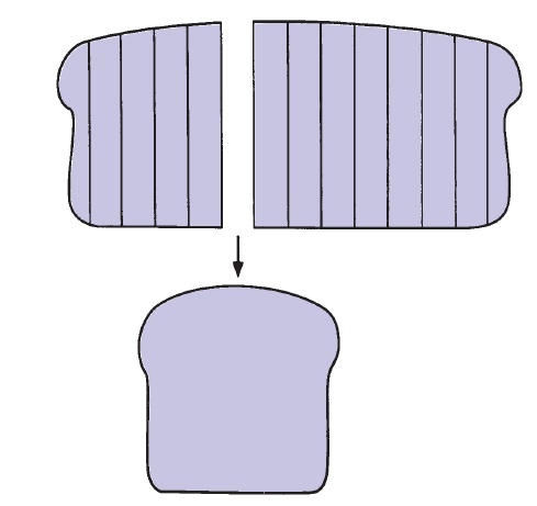
CT involves sectional anatomy imaging or anatomy in the sagittal, coronal, and axial (cross-sectional, transverse) planes. CT imaging is best understood if the anatomic site to be examined is thought of as a loaf of sliced bread; an image of each slice of bread is created without imaging the other slices. This is in contradistinction to a radiograph, which captures the whole loaf of bread as in a photograph.
CT Scanner parts and mechanics
- Parts of CT scanner includes,
- X-ray tube gantry (round opening)
- X-tube
- Collimator and detector
- Computer controlled patient couch
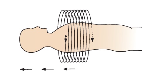
- CT images are produced by a combination of x-rays, computers, and detectors. A computer-controlled couch transfers the patient in short increments through the opening in the scanner housing. In the original, now near-extinct standard CT unit, the x-ray tube located in the housing (gantry) rotates around the patient, and each anatomic slice to be imaged is exposed to a pencil-thin x-ray beam.
- Same like in x-ray radiograph, the amount of the x-ray beam that passes through each slice or section of the patient will be inversely proportional to the density of the traversed tissues.
- The x-rays that pass completely through the patient eventually strike detectors (not film), and the detectors subsequently convert these incident x-rays to an electron stream.
- This electron stream is digitized or converted to numbers referred to as CT units or Hounsfield units; then computer software converts these numbers to corresponding shades of black, white, and gray.

Thickness of images / slices
The thickness of these axial images or slices can be varied from 1 to 10 mm depending on the indications for the study. For example, in the abdomen and lungs we commonly use a 10-mm slice thickness because the structures are large.
Types of CT
1. Helical or spiral CTThis technology is more similar to standard CT. In helical or spiral CT, the patient continuously moves through the gantry while the x-ray tube continuously encircles the patient.
2. Multi-slice /Dynamic CT
This equipment has multiple contiguous rows of detectors that yield multiple tomographic slices with only one rotation of the x-ray tube around the patient. Remember that tradition CT can produce only single image with single rotation.
3. Dual-Source CTDual-energy scanners also utilize multiple detectors and helical scanning. It utilizes two energy sources from single x-ray tube. Two energies are low and high energy x-rays. It has some advantages and some disadvantages. That is, an image generated with low- and high-energy x-rays will have different gray scale values for the same object. The two images resulting from the low- and high-energy x-rays can be combined using a weighted subtraction. Dual-energy imaging has a number of applications including direct removal of bone for angiographic imaging, plaque characterization, lung perfusion, identification of ligaments and tendons, and assessment of tissue composition. Radiation dose is a potential concern or disadvantage of using dual-source scanners.
Contrast media
CT imaging can be taken with or without intravenous contrast media. Contract media enhances the visualization of blood vessels, organs and tumours. It allows clinician to distinguish between anatomy and pathological process.
Indications of CT
- Trauma
- Intracranial hemorrhage (suspected or known)
- Abdominal injury, especially to organs
- Fracture detection and evaluation
- Spine alignment
- Detection of foreign bodies (especially in joints)
- Diagnosis of primary and secondary neoplasms (liver, renal, brain, lung, and bone)
- Tumour staging.
SHARE:


- Carol A. Boles, MD, William E. Erkonen, MD et al., Radiology - the basics and fundamentals of imaging., fourth edition.
- Sutton D., textbook of radiology.
- Michael Y.M. chen et al., Basic radiology., second edition.
| Name | : | Deva senathipathi |
| Qualifications | : | Physiotherapist |